
Image restoration
Image restoration refers to the recovery of an original signal from degraded observations.Image restoration is different from image enhancement in that the latter is designed to emphasize features of the image that make the image more pleasing to the observer, but not necessarily to produce realistic data from a scientific point of view. Image enhancement techniques (like contrast stretching or de-blurring by a nearest neighbor procedure) provided by "Imaging packages" use no a-priori model of the process that created the image.
With image enhancement, noise can be effectively removed by sacrificing some resolution. However, this is not acceptable in many applications as no distinction can be made between noise and real signal. In a Fluorescence Microscope the resolution is bad as it is. More advanced image processing techniques must be applied to improve the resolution to recover the object.
Deconvolution is an example of an image restoration method. It is capable of:
- Increasing resolution, especially in the axial direction.
- Removing noise.
- Increasing contrast
Since axial imaging performance is the prime reason for researchers to invest in expensive optical equipment like confocal or two-photon excitation microscopes, the capability of increasing axial resolution with 'merely' a deconvolution software technique has considerable value.
In image restoration the information provided by the microscope is only taken as indirect evidence about the object. By itself the image needs not even to be viewable.
- A microscopy image contains more information than is visible in the image.
- Often, details are hidden in the noise, out-of-focus signal, or masked by other features.
- Artifacts may confuse the viewer.
- Information may be present in implicit form so it can only be retrieved with the addition of a-priori knowledge.
Examples
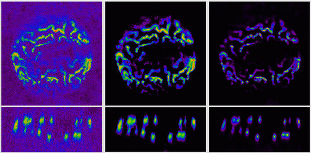
Chromosomes in a sea urchin nucleus recorded with a confocal microscope. Left: raw image data. Middle: filtered and enhanced data. Right: after image restoration. Top row: XY plane at z = 38; bottom row: XZ plane at y = 229. Image dimensions: 22.5 × 22.5 × 11.4 µm. Data courtesy Prof. Thomas Baechi, University Zurich, Switzerland.
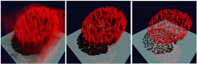
Chromosomes in a sea urchin nucleus recorded with a confocal microscope. Left: Simulated Fluorescence Process (SFP) rendering of the raw image after thresholding the background. Middle: SFP rendering after applying a 3D gaussian noise filter and a Soft Threshold. Right: SFP after applying image restoration with the Huygens Software. Data courtesy Prof. Thomas Baechi, University Zurich, Switzerland.
See other examples in Restoration Examples.